Experimental animals
Figure 1 illustrates the experimental and workflow of FKN detection. WKY rats were purchased from Changsha Tianqin Biotechnology Co., LTD., Hunan. Myeloperoxidase (MPO, Sigma-Aldrich, M6908-5UN), Freund’s complete adjuvant (Sigma-F5881), MPO-ANCA-IgG ELISA Kit (CB-E08675R; Cusabio Ltd., Wuhan, China), Anti-rat Fractalkine neutralizing antibody (AF537; R&D Systems), Fractalkine ELISA kit (Bethyl Laboratory), Antibodies against Fractalkine ( DF12376, Affinity Biosciences, Changzhou, China). Anti-glyceraldehyde3-phosphatedehydrogenase (GAPDH; AF7021, Affinity Biosciences, Changzhou, China). Creatinine(Cr)Colorimetric Assay Kit (Sarcosine Oxidase Method, E-BC-K188-M, Elabscience Biotechnology Co.,Ltd, Wuhan, China; Urea (BUN) Colorimetric Assay Kit (Ur, ease Method, E-BC-K183-M, Elabscience Biotechnology Co.,Ltd, Wuhan, China); Urine Protein Colorimetric Assay Kit(E-BC-K252-M, Elabscience Biotechnology Co.,Ltd, Wuhan, China).
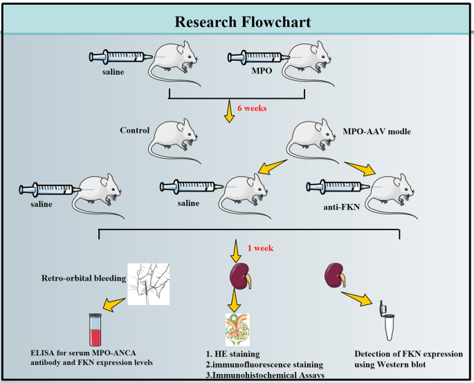
Schematic illustration of the experimental procedures. Myeloperoxidase(MPO), myeloperoxidase and anti-neutrophil cytoplasmic antibody associated vasculitis (MPO-AAV), anti-fractalkine (anti-FKN)
Establishment of animal models and collection of tissue specimens
Thirty Wistar-Kyoto (WKY) rats were randomly divided into three groups (n = 10/group) the control group; the MPO-AAV group in which rats were given a one-time intraperitoneal injection of 400 µg/kg MPO (MPO dissolved in sterile water for injection at a concentration of 500 µg/ml, then mixed with an equal amount of Freund’s complete adjuvant [23,24,25,26,27]; and the MPO-AAV + anti-FKN group, in which rats were given a single intraperitoneal injection of 400 µg/kg MPO for 6 weeks, then anti-FKN (1 µg/ rat /day, i.p) [28, 29]. One week later, orbital blood was collected on EDTA-K2 anticoagulant tube, and the rats were killed by disjoint method. The abdominal cavity was fully exposed, both kidneys were removed, half kidney tissue was cut open, half kidney tissue was fixed with 10% formalin, paraffin embedding was used for immunofluorescence staining and HE staining, and the other half kidney tissue samples were extracted and stored at -80℃ for total protein extraction.
Detection of renal function indicators
According to related kit instructions, urinary albumin (UAER), serum creatinine (Scr) and blood urea nitrogen (BUN) contents in urine were detected by automatic biochemical analyzer.
Enzyme-linked immunosorbent assay
The levels of MPO-ANCA, and FKN in rat serum were detected using the respective enzyme-linked immunosorbent assay kits. The absorbance was measured at 450 nm using a microplate reader.
Western blot assay
The total protein of rat kidney tissue was extracted based on previous research methods [30], the protein concentration was detected by BCA method, the protein sample was added for SDS-PAGE electrophoresis, the membrane was transferred at a constant voltage of 100 V for 110 min, the protein was transferred to the nitrocellulose membrane, and the sealing liquid was used for 60 min. Anti-FKN (DF12376), 1:1100) (Affinity Biosciences, Changzhou,, China) was added and incubated at 4℃ overnight. After Tris-HCI Tween(TBST) washing for 3 times, corresponding horseradish peroxidase labeled secondary antibody was added (1: 2000), incubated in a shaker at room temperature for 50 min, enhanced chemiluminescence (ECL) development was performed. ImageJ software was used for semi-quantitative analysis, and the relative content was represented by the ratio of gray values of target protein bands to GAPDH protein bands. The experiment was repeated three times.
Immunofluorescence staining
Paraffin sections of kidney tissue were fully dewaxed by xylene, 100%-75% gradient alcohol to water, then boiled in EDTA buffer solution (pH8.0) for 20 min, washed 3 times in PBS after natural cooling, and incubated at room temperature for 1 h in the closed solution (containing 2% BSA and 10% goat serum). Then FKN monoclonal antibody was incubated overnight, followed by PBS for 3 times on the second day, and the corresponding fluorescent secondary antibody was incubated for 1 h. After PBS for 3 times, DAPI was incubated for 10 min and nucleated. After PBS cleaning, the tablets were sealed with anti-fluorescent attenuation tablets. The FV3000 laser scanning confocal microscope was used to obtain images of 5 fields per rat.
Hematoxylin-eosin staining
The renal tissue sections were stained by Thermo scientific GeminiAS, sealed by Clearvue automatic slice sealing machine, scanned by 3DHistech Pannoramic Midi scanner, and collected by caseviewer software.
Immunohistochemical assays
Single-labelled immunohistochemical assays were performed using p65NF-κB and IL-6 primary antibodies. Sample slides were incubated with the corresponding primary antibodies overnight at 4 °C, then labelled with secondary antibodies (goat anti-rabbit, PV-6000, ready-to-use, ZSBG-BIO, China) for 20 min at 37 °C. PBS was rinsed, and the labelled tissues were labelled with freshly prepared DAB chromogenic reagent. The reaction time was observed with a microscope until the positive expression showed brownish yellow colour. The nuclei were stained with hematoxylin staining solution (BA-4041, BASO, China). The slides were then treated with differentiation solution (C0163M, Beyotime, China) for a few seconds and washed with counterblue in running tap water. As identified by DAB reagent (ZLI-9018, ZSBG-BIO, China), the cells of positive cells were brownish-yellow in colour and the nuclei were blue.
Statistical analysis
SPSS 23.0 statistical software was used for data analysis. All measurement data were expressed as X ± S, and data were analyzed by means comparison of multiple samples and pair comparison. α = 0.01 was used as the test level, and the homogeneity test of variances was conducted. P < 0.01 was considered statistically significant.
- The Renal Warrior Project. Join Now
- Source: https://bmcnephrol.biomedcentral.com/articles/10.1186/s12882-024-03565-3