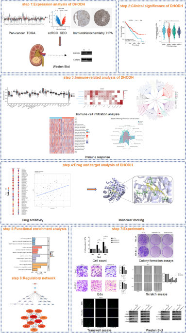
DHODH gene information
We used the GeneCards database20 (https://www.genecards.org/) to visualize the chromosome and subcellular locations of gene encoding DHODH. The protein topology of DHODH was obtained from PROTTER21 (https://wlab.ethz.ch/protter/start/). The RNA expression levels of the DHODH gene in 20 normal tissues were obtained from the NCBI database (https://www.ncbi.nlm.nih.gov/).
Data collection and preprocessing
RNA-seq data processed using the STAR pipeline from 33 tumor projects were downloaded and extracted in TPM format from TCGA database (https://portal.gdc.cancer.gov). RNA-seq data processed using the Toil pipeline22 from TCGA, and GTEx in TPM format were obtained from UCSC XENA (https://xenabrowser.net/datapages/). The corresponding TCGA data for pan-cancer and GTEx data for normal tissues were collected. The ccRCC datasets, GSE1260623 and GSE18933124, were obtained from the GEO database (https://www.ncbi.nlm.nih.gov/geo/).
To validate the expression of DHODH in pan-cancer, the TIMER database25 (http://timer.cistrome.org/) was used to visualize the differential expression of the DHODH gene across various cancer types. The effect of DHODH on pan-cancer outcomes was assessed using the TIMER database.
DHODH expression
Using the Stats and Car packages in R (version 4.2.1), Wilcoxon rank-sum test analysis was conducted to examine the differential mRNA expression of DHODH across all cancers. Differences in the mRNA expression levels of DHODH were also assessed in different disease states (tumor or normal) and cancer stages (including TNM staging and histological grading) of ccRCC, and visualized using box plots, violin plots, and paired sample line plots with the ggplot2 package. The data were standardized using the normalizeBetweenArrays function in the Limma package. Differentially expressed genes (DEGs) were selected based on the criteria: “|Log2(Fold Change)|≥ 1 and P value < 0.05.” Volcano plots were used to display the expression of DHODH in the GSE12606 dataset. The specific antibody against DHODH, HPA-010123, was obtained from the Human Protein Atlas (HPA, http://www.Proteinatlas.org/) online database26 for immunohistochemical staining of clinical ccRCC and normal kidney tissues.
Kaplan–Meier plot analysis
By employing the survival package, Kaplan–Meier survival analysis and log-rank tests were performed using TCGA-KIRC dataset to investigate the relationship between gene expression and overall survival (OS), progression-free interval (PFI), and disease-specific survival (DSS) in patients with ccRCC. Subgroup survival analyses based on TNM stage and histological grade were also conducted, and the results were visualized using the Survminer and ggplot2 packages.
Immune cell infiltration analysis
Using the single-sample Gene Set Enrichment Analysis (ssGSEA) algorithm from the GSVA package (version 3.6)27, immune infiltration analysis was conducted using TCGA-KIRC dataset to assess the enrichment status of 24 immune cell types28 across samples grouped by the expression levels of DHODH. The Wilcoxon rank-sum test was employed for this analysis. Thereafter, Spearman correlation analysis was performed to determine the effect of DHODH expression on immune cell infiltration, yielding Spearman correlation coefficients. The results were visualized using the Laplace method and grouped into violin plots. A focused examination was performed on the Spearman correlations between DHODH expression and the infiltration levels of six pivotal immune cell types (B cells, CD8 + T cells, macrophages, neutrophils, NK cells, and dendritic cells) using the circlize package; the results were displayed in chord diagrams.
The TISCH database was used to explore the single-cell expression of DHODH, and the TIMER 2.0 database was employed to determine the effect of DHODH expression on tumor immune cell infiltration in 115 different subtypes of tumors. The results are displayed in a circular clustering diagram.
The effect of the expression level of DHODH on the anticancer immune status in ccRCC was analyzed using the Tracking Tumor Immunophenotype (TIP) database29 (http://biocc.hrbmu.edu.cn/TIP). The immune activity scores between samples with high and low DHODH expression were compared using Student’s t-test or Wilcoxon rank-sum test, and the results were visualized using the R packages, pheatmap and ggplot2. To comprehensively illustrate the anticancer immune status of samples with high and low DHODH expression, all samples were divided into high- and low-expression groups based on the median expression value of DHODH, and samples with expression values close to the mean value of each group were selected as examples (low-expression sample: TCGA-CW-6090-01A-11R-1672-07; high expression sample: TCGA-CZ-5453-01A-01R-1503-07). Thereafter, the effect of DHODH on the seven steps of antitumor immune response was analyzed. The correlation between DHODH expression and the immune checkpoint were analyzed using scatter plots30.
Drug sensitivity analysis and molecular docking
To analyze small-molecule drugs targeting DHODH, the GSCALite online database31 was used to calculate the Spearman correlation coefficient to determine the correlation between DHODH expression levels and the drug sensitivity (50% inhibitory concentration (IC50)) of 65 small-molecule drugs from the GDSC and CTRP databases.
RNA expression data (RNA: RNA-seq) and drug data (compound activity: DTP NCI-60) of the NCI-60 cell lines were downloaded from the CellMiner database. The correlation between DHODH and small-molecule drugs was reanalyzed and presented as a dot plot. The sensitivity of small-molecule drugs in the DHODH high- and low-expression groups is displayed using a box plot.
For molecular docking, the name, molecular weight, and 3D structure of the small-molecule drugs were determined using the PubChem database, and the 3D structure corresponding to the DHODH gene was downloaded from the RCSB PDB database (https://www.rcsb.org/). Ligands and proteins were prepared for molecular docking using the AutoDock Vina software (http://vina.scripps.edu/). Finally, the AutoDock 1.5.6 software was used to dock the structure of DHODH with that of the small-molecule drug, where the affinity (kcal/mol) value represented their binding ability. The lower the affinity (kcal/mol), the more stable the binding between the ligand and receptor. The results were visualized using the PyMOL software.
Single-gene differential expression and enrichment analysis
The statistical ranking of genes with DHODH expression higher or lower than the median was defined as the high or low DHODH expression groups, respectively. The DEGs between these two groups were identified using the DESeq2 R package32 and an unpaired Student’s t-test. Genes with adjusted P value < 0.05 and |Log2(Fold Change)|≥ 1 were considered statistically significant and were included in the subsequent analysis. All DEGs are presented in volcano plots and heat maps.
To determine the function of DHODH, the DEGs were subjected to Gene Ontology (GO)33 and Kyoto Encyclopedia of Genes and Genomes (KEGG)34 analyses using the ClusterProfiler package35 in the R software. Statistical significance was set at P < 0.05. GO terms were divided into three categories: biological processes (BP), cellular components (CC), and molecular functions (MF). Some results were visualized using bar plots generated using the ggplot2 package.
Gene set enrichment analysis (GSEA)36 was performed for all DEGs with statistical significance. This analysis aimed to identify differences in functional phenotypes and signaling pathways between the high- and low-expression groups. The ClusterProfiler package was used for this analysis, and the reference gene set was obtained from the c2.cp.all.v2022.1. Hs.symbols.gmt gene set database within MSigDB Collections. P < 0.05 and normalized enrichment score (|NES|) > 1 indicated significant enrichment.
Identification of the ceRNA and protein interaction (PPI) networks
To explore the potential molecular mechanisms of DHODH in ccRCC, StarBase (https://starbase.sysu.edu.cn/) and mirRTarBase (https://mirTarBase.cuhk.edu.cn/) were used to predict the miRNAs that can interact with DHODH and lncRNAs that can bind to miRNAs, respectively. To better understand the role of DHODH in ccRCC resistance, we compared the expression levels of miRNAs between sunitinib-resistant samples and sunitinib-sensitive samples in the GEO dataset, GSE189331, and selected differentially expressed miRNAs that were negatively correlated with DHODH expression (logFC < − 1 and P < 0.05). Furthermore, based on TCGA-KIRC, Kaplan–Meier analysis was performed to identify prognostic lncRNAs in ccRCC and Cytoscape (version 3.10.0) was used to display the interactions among mRNAs, miRNAs, and lncRNAs to analyze the potential ceRNA regulatory mechanisms involved in ccRCC.
The STRING database (http://string-db.org) (version 12.0)37 was used to identify proteins that interact with DHODH. Thereafter, a PPI network with complex regulatory relationships was constructed. Interactions with medium confidence > 0.4 were considered statistically significant. The MCODE plugin in Cytoscape was used to analyze key functional modules. The selection criteria were as follows: K-core = 2, degree cutoff = 2, maximum depth = 100, and node-score cutoff = 0.2. Subsequently, KEGG and GO analyses were performed using these shortlisted genes.
Cell culture and RNA interference
The human RCC cell lines, 786-O and OS-RC-2, were obtained from Procell Life Science&Technology Co., Ltd. (Procell, Wuhan, China) and cultured in Procell’s basic medium (ROMI-1640, Procell, Wuhan, China) supplemented with 1% penicillin/streptomycin (MA0110, Meilunbio, Shanghai, China) and 10% fetal bovine serum (TIANHANG, Zhejiang, China). The 786-O and OS-RC-2 cells were maintained at 37 °C in a 5% CO2 environment.
DHODH-targeting siRNAs (hDHODH-259, hDHODH-849) were transfected into 786-O and OS-RC-2 cells for 48 h using the DharmaFECTTM1 transfection reagent kit (4000-3, Engreen Biosystem Co. Ltd.), according to the manufacturer’s instructions. The siRNAs targeting DHODH and scrambled siRNAs were obtained from Sangon Biotech (Wuhan, China).
Cell proliferation and migration assays
First, 786-O and OS-RC-2 cells (10 × 104 cells/well) were seeded in 6-well plates and cultured for 48 h. RNA interference (RNAi) technology was used to interfere with the expression of DHODH in 786-O and OS-RC-2 cells. On days 3 and 5 of cell culture, the 786-O and OS-RC-2 cells were digested with trypsin. The proliferation of 786-O and OS-RC-2 cells was detected using the Neubauer counting chamber cell counting method.
The EDU proliferation assay was performed using the BeyoClick™ EDU-488 Cell Proliferation Assay Kit (C0071S, Beyotime, Shanghai, CHN). Briefly, cells were seeded (3 × 10⁴ cells/well) in a 6-well plate containing the corresponding concentration of EDU reagent (1:500) for 3 h. The cells were then washed twice with 1 × PBS (BL302A; Biosharp, Anhui, CHN) for 5 min, incubated with 4% paraformaldehyde for 30 min, permeabilized with 0.3% Triton X-100 immunostaining permeabilization solution (BL935A; Biosharp, Anhui, CHN), and staining using the reaction solution. Images were captured using a fluorescence microscope.
The effect of DHODH on the colony forming ability of 786-O and OS-RC-2 cells was evaluated. 786-O and OS-RC-2 cells (500 cells per well) were seeded in 6-well plates, cultured for 1 week, and subjected to RNAi-mediated interference of DHODH expression. Cells were cultured for another week. The resulting colonies were fixed with 4% paraformaldehyde and stained with crystal violet solution.
For the wound healing assay, healthy 786-O and OS-RC-2 cells were seeded in 6-well plates (50 × 104 cells/well). After 48 h culture, the RNAi technology was used to interfere with the expression of DHODH in 786-O and OS-RC-2 cells. A scratch was mechanically created using a 1 ml sterile pipette tip, and the cells were cultured for 24 and 48 h. Images were captured at 0, 24, and 48 h, and the wound healing rate was calculated using the ImageJ software.
For the transwell assay, cells were seeded (20 × 104 cells/well) and cultured for 48 h. RNAi technology was used to interfere with the expression of DHODH in 786-O and OS-RC-2 cells. The 786-O and OS-RC-2 cells were digested with trypsin, and a cell suspension containing 2 × 10⁴ cells in 300 (mu)L of complete culture medium containing 2% fetal bovine serum was seeded in the upper chamber. The lower chamber was filled with complete culture medium containing 10% fetal bovine serum. After 24 h culture, the cells were fixed with 4% paraformaldehyde, stained with crystal violet, and observed and quantified using ImageJ software to measure their invasion ability.
Clinical tissue samples
Tumor tissues and the corresponding adjacent normal tissues were obtained from five patients with primary ccRCC who were admitted to the Urology Department of Zhongshan Hospital of Xiamen University in 2023. All patients with CRC underwent curative surgery, and their pathological diagnosis was ccRCC without other malignant tumors. None of the patients underwent preoperative radiotherapy, neoadjuvant chemotherapy, or other treatments. All patients signed a written informed consent form. The study was approved by the Medical Ethics Committee of Zhongshan Hospital of Xiamen University (2024-553). The study was conducted in accordance with the principles of the Declaration of Helsinki. All methods were performed in accordance with the relevant guidelines and regulations.
Western blot analysis
Total protein was extracted from cells or tumor tissues using RIPA lysis buffer (BL504A; Biosharp, Anhui, CHN) supplemented with protease inhibitors and EDTA (Beyotime, Shanghai, CHN). Protein amounts were quantified using a BCA Protein Assay Kit (P0010; Beyotime, Shanghai, CHN). Equal amounts of protein were separated by performing SDS-PAGE (Beyotime, Shanghai, CHN) using on 10–12% resolving gels and were transferred onto a 0.2 μM PVDF membrane (Immobilon®-PSQ, Ireland). The membranes were blocked with 5% skim milk and incubated with the following primary antibodies: DHODH (14877-1-AP, Proteintech), N-cad (66219-1-lg, Proteintech), Vimentin (E-AB-18212, Proteintech), Snail (26183-1-AP, Proteintech), MMP2 (E-AB-40409, Proteintech), Parp1 (13371-1-AP, Proteintech), GPX4 (67763-1-lg, Proteintech), SLC7A11 (26864-1-AP, Proteintech), FTH1 (10727-1-AP, Proteintech), and GAPDH (60004-1-lg, Proteintech), diluted 1:1000 in Western primary antibody dilution solution (E-IR-R125, Elabscience, Wuhan, CHN). Thereafter, the membranes were incubated with the corresponding secondary antibodies, rabbit anti-mouse (SA00001-2-lg; Proteintech) and goat anti-rabbit (SA00001-2-lg; Proteintech), diluted 1:3000. The signal was detected using TanonTM Femto-sig ECL Western Blotting Substrate (180-506, Tanon).
Expression of ferroptosis-related protein in renal tissue based on immunohistochemistry and transmission electron microscopy (TEM)
Conventional ccRCC tissues and normal renal tissues adjacent to the carcinoma were embedded in paraffin and sliced. The sliced tissues were incubated with 3% hydrogen peroxide at room temperature for 15 min and washed 3 times with PBS (3 min/wash). Following removal of the excess water, the tissues were incubated with the diluted primary antibodies, including those against FTH1, GPX4, and SLC7A11 (1:300, the product catalog number is the same as that for the western blotting experiment) overnight at 4 °C. Immunoreactive bands were stained according to the instructions of the immunohistochemical kit (Beijing Zhongshan Jinqiao Biotechnology Co., Ltd., PV-9000) and concentrated DAB kit (Beijing Suolaibao Technology Co., Ltd., DA1010).
After pre-treatment, cells were collected, precipitated, and fixed with 2.5% glutaraldehyde for 2 h at 4 °C. Images were captured after tissue dehydration, infiltration, slicing, embedding, and staining.
Data analysis
The experimental data were analyzed using GraphPad Prism 9.3.0 software. The results are presented as mean ± standard deviation (mean ± SD). One-way analysis of variance (ANOVA) was used for multiple comparisons between groups. A P < 0.05 indicated statistical significance.
- The Renal Warrior Project. Join Now
- Source: https://www.nature.com/articles/s41598-024-62738-0
